They are so small that thousands of them could pack into the dot
of this i. Yet despite their size, proteins are the building blocks of life.
They are vital to just about every biological process you can name: sleeping
and eating, working and playing, even laughing and crying.
At the cellular level, proteins are in charge of everything from gene expression
to what materials can enter or exit a cell. Because proteins are essential
to nearly every living process, scientists want to learn everything they can
about them. And because proteins are so small (they are measured by the angstrom,
the equivalent of one-hundred millionth of a centimeter), highly refined techniques
like X-ray crystallography are required to "see" them.
Knowing a protein's physical attributes - what it actually looks like - helps
scientists understand how it functions in relation to other molecules in the
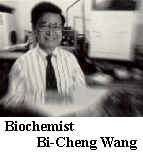
cell, said biochemist Bi-Cheng Wang, the UGA Eminent Scholar of X-ray Crystallography
and professor of biochemistry.
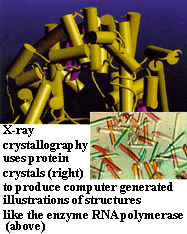
If you've never heard of X-ray crystallography, you're in the majority. Basically,
it's a magnifying technique that uses X-rays to bombard crystals - in this
case, proteins that have been grown
as crystals - then capture their interference patterns on light-sensitive material.
Scientists then analyze these patterns with high-speed computers that draw
amazingly accurate pictures of molecular structures.
Once they know a molecule's structure based on X-ray crystallography, scientists
can better predict how the molecule works - whether its job is to copy the
genetic information from DNA, turn genes off or on, speed up chemical reactions,
identify viruses or other foreign substances, or simply cart materials in and
out of the cell. Knowing the three-dimensional structure of a protein also
can help explain how it identifies a precise sequence on a strand of DNA. This
kind of information helps advance science on many fronts: genetics, medicine
and drug design, to name a few.
"It is important to know a protein molecule's structure because it will
show you how it will function physically," Wang said. "Anything that
functions in nature has something to do with its structure. It will function
a certain way because it has a certain structure. This applies to everything.
And once we understand a structure we can control its function."
Because molecules in a crystal are aligned in an orderly fashion, they yield
very predictable interference or diffraction patterns that are related to
the spacing of atoms in the molecule and therefore unique to the crystal's
structure. For instance, X-ray crystallography can help explain everything
from how a drug can be designed to be more effective in the human body to
why diamond crystals are extremely hard while salt crystals dissolve in water.
"The magnification of X-ray crystallography is considerably greater than
electron microscopy or any other technique currently available," Wang said. "We
can determine resolution in terms of two or three angstroms, and that enables
us to study large biological molecules like proteins, DNA and viruses."
"Only on special occasions can we actually get data to the resolution that
we can see individual atoms. But we can see the gross shape of each amino acid
in the protein," said John Rose, a UGA associate research biochemist who
has worked with Wang for 16 years.
Fundamentals of Life
Of key interest to Wang, Rose and their colleagues is an enzyme called RNA
polymerase that helps transcribe discreet sections of the genetic code from
DNA to RNA. The RNA then carries a copy of the genetic blueprint on how to
make a specific molecule - an enzyme, a hormone or perhaps a disease-fighting
antibody - to an assembly site in the cell.
"This enzyme interprets genetic information," Wang said. "And
that is a very fundamental and important life process."
In 1993, while still on the faculty of the University of Pittsburgh, Wang
was the senior researcher who determined the crystal structure of an RNA
polymerase isolated from a virus. The discovery was considered so important
that it was included in the 1995 edition of the Encyclopedia Britannica.
Knowing the structure of RNA polymerase helps scientists understand how genetic
information is decoded at the atomic level.
Funding from the National Institutes of Health (NIH) and the Georgia Research
Alliance is helping Wang's team refine their technique to get an even greater
magnification of the enzyme's structure. Key to achieving that goal is having
access to state-of-the-art equipment like their new charged couple device (CCD)
X-ray detector, which Rose said "is probably the first one installed in
a university lab anywhere in the United States, or the world, for that matter."
With increased magnification comes a clearer understanding of how RNA polymerase
precisely identifies and physically attaches to a DNA strand. Such information
will shed light on the mechanics of how DNA directs cell growth, development
and replication.
"Now that we have determined the structure, we are extending the resolution
of the molecules from 3.3 angstroms to 2.6 angstroms," Wang said. "We
are learning much more detail about the atomic arrangement but it takes a lot
of effort just to extend it that much. First you have to get a better crystal."
Growing the Perfect Crystal
A good protein crystal for X-ray crystallography has a very orderly molecular
arrangement, which increases the resolution and provides a much better picture
of its structure.
To grow an orderly crystal can take anywhere from a few days to several months.
And that's after completing an often lengthy process to purify the protein
in the first place.
"Preparing crystals is one of the most important parts of crystallographic
studies," Wang said. "You have to be careful and very patient, especially
because you do not know if you will get a crystal."
To see if a protein will form a crystal, the researchers perform a quick screening
test. If that is successful, they will try to grow larger crystals - about
the size of a pencil point - using one of two processes. One method is a sophisticated
variation of the same evaporation technique you may have used to grow rock
candy. The other is a diffusion method that uses a membrane that lets smaller
molecules pass into a chamber but blocks larger ones. Either way, the like
protein molecules in the solution begin to pack together to form an orderly
crystal.
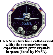
Their quest for a better crystal has led Wang and Rose to try growing crystals
in space aboard the space shuttle Columbia and the Mir space station. They
are collaborating with scientists at the George C. Marshall Space Flight Center
in Huntsville, Ala., and the University of Alabama in partnership with the
U.S. Microgravity Lab II. Growing crystals in space eliminates the pull of
gravity and allows the crystals to grow with less interference.
Of the three proteins grown aboard the Columbia, only the RNA polymerase, which
undergoes a complicated crystallization process on Earth, failed to produce
good crystals. The other proteins B a hormone vasopressin-protein complex and
a newly discovered liver growth factor B actually grew better in space than
on Earth.
"The RNA polymerase sample did not grow because there simply was not enough
time," Wang said. "So we repeated it on the Mir, which was in space
for six months, to see if it would grow given more time."
Again, the results were disappointing but the researchers say they believe
that was due, in part, to technical problems, including variations in temperatures
during the experiment. A new batch of crystal experiments was carried to the
Mir aboard the same shuttle that brought astronaut Shannon Lucid back to Earth,
but Wang and Rose will not know the results of this latest vasopressin effort
until some time this spring.
Crystal-Clear Hormones
The space-grown vasopressin is among the dozen hormones associated with a group
of brain proteins called neurophysins that are targeted for Wang's X-ray studies.
With grants from NIH and the Pittsburgh Super Computer Center, Wang's team
is working with Esther Breslow, a professor at Cornell University Medical College,
to understand how structural characteristics of these hormones influence their
functions, especially in relation to the neurophysins that always seem to be
coupled with them.
For example, the hormones vasopressin and oxytocin are found in high concentration
in the brain's pituitary gland and are always complexed in a one-to-one ratio
with neurophysins. Although the two hormones influence very different physiological
processes - vasopressin affects water balance and blood pressure and also may
improve memory during early aging while oxytocin affects milk secretion and
uterine contraction - Wang's group discovered they have similar structures.
Their similarity has intrigued scientists because of the recent discovery that
these two hormones also play surprisingly similar roles in orchestrating social
relationships: such as the love between a man and a woman, including sexual
behavior, as well as the bonding between parents and children. In fact, oxytocin
also is called the cuddle compound or love hormone.
Armed with structural information on these hormones, scientists may be able
to clarify the physiological connection between love and hormones. For example,
the structures help explain how molecules of oxytocin and neurophysin are packed
together in the nerve endings in very high concentrations in such a way that
they can be retrieved very quickly.
"When you think about it, love somehow is a biological process," Wang
said. "There has to be some kind of hard wiring in the brain. We think
the neurophysins help store the oxytocin in the nerve endings. It's almost
like the orderly storage you would find in a warehouse that enables you to
retrieve items very quickly. If the body needs to respond quickly, it can't
say, 'Wait. I have to synthesize this and it"ll take a day or two.'"
So far, the team has crystallized the 12 neurophysin-hormone complexes and
determined nine of their structures. When they first started this study in
1978, the researchers simply were interested in why the hormones and neurophysins
have such a high affinity.
"Now we know that a knowledge of the structure-function relationship of
these [hormones] also may be significant in understanding the molecular basis
of emotions as well as problems related to aging, mental illness and other areas
of endocrinology," Wang said.
Crystals and Medicine
X-ray crystallography also is lighting the path to better medicine by showing
how cancer invades cells or why drug therapies work. For instance, it can help
scientists understand how viruses physically invade and infect cells.
Wang's team has successfully grown crystals of some enzymes that protect against
certain cancer-causing agents and that also may account for drug resistance
in some tumor cells. Experiments have shown that theses enzymes from the glutathione
S-transferase (GST) system also may be involved in life span and aging. So
far, the team has grown 13 forms of the GST enzymes and has determined structures
for six of them.
"A knowledge of the structure-function relationship of GSTs may have long-term
significance in understanding problems related to cancer prevention, tumor control
and parasitic diseases," Wang said. It also will increase our knowledge
of how large molecules of the same type recognize each other.
The UGA researchers also are pointing their X-ray beams at a recently isolated
liver protein, augmentor of liver regenerator (ALR). Unlike other organs in
the human body, the liver has the ability to regenerate after it is damaged.
ALR, which is a key to this regenerative process, is found in the liver only
during development stages, such as during fetal growth or following surgical
removal of a section of the liver, Rose said.
"That's the interesting part," he said. "It doesn't cause the
liver to regenerate but it enhances the regeneration that's occurring. Perhaps
more of a diseased liver could be removed and still have it regenerate by supplementing
the ALR already in the liver."
Although they have not yet figured out ALR's structure, they believe its structure
may open new doors to treat hepatitis, liver cancer and other liver diseases,
Wang said. Scientists with the Thomas E. Starzl Transplant Institute, led by
liver transplant surgeon Thomas Starzl and professor Antonio Francavilla, isolated
ALR and have purified about 50 milligrams, which must be stretched to fuel
several research projects, including limited testing in patients.
"They gave us two milligrams about two years ago for us to start the project,
and we are hoping we will have determined the structure in the very near future," Rose
said.
"Knowing the structure is the first step to knowing how this protein functions
and helps regenerate a cell," Wang said. "And that could also help
someday in designing a drug to help regenerate a damaged liver, for example."
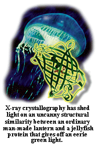
Green fluorescent protein (GFP) also has captured the attention of the UGA
X-ray crystallographers. Found naturally as a protein in the jellyfish Aequorea
victoria that gives off a green light, GFP is used in medicine and research
to track proteins and perform diagnostic tests. For instance, if you want to
know where a certain protein is made in the cell, you can mark it with a fluorescent
probe of GFP and see where it lights up the cell.
Wang and a host of other researchers from the University of Georgia, the University
of Pittsburgh, the Scripps Institution of Oceanography, the University of California
at San Diego and the University of Oregon recently determined GFP's three-dimensional
structure. Surprisingly, it turns out that GFP looks like a lantern: The light-emitting
element is in the center, surrounded by a protective protein shield.
"This is a new type of structure and the location of the light-emitting
element in the center of the shield makes perfect structural sense in terms of
its function," Wang said.
The UGA researchers also have recently determined the structure of the enzyme
aldehyde dehydrogenase. This could have medical application because the enzyme
helps convert alcohol to simpler substances which the body can then eliminate.
In the process of determining the structure of aldehyde dehydrogenase, the
researchers discovered a previously unknown mechanism for an enzyme helper
called NAD. At first the researchers thought they had made a mistake somewhere.
After checking and rechecking every procedure and calculation, they determined
their data was right, and that opens up all kinds of new possibilities in the
lock-and-key field of how molecules recognize each other and attach in very
specific ways.
"X-ray crystallography is regarded as the absolute [authority] right now
in terms of defining structure," Rose said. "If you say the distance
from this atom to this atom is a certain length, people will take you at your
word and that puts a lot of pressure on us. When we publish our findings we know
we have to be correct [because] when you say something in this field, it becomes
the gold standard."
For more information, access http://www.uga.edu/~biocryst/.
Judy Bolyard Purdy is UGA's director of research communications and editor
of Research Reporter. A former naturalist with the National Audubon
Society, she has published a book on medicinal plants and has degrees in biology,
botany and journalism.