

“Cat Scan” Reveals
Brain Tumor
By Rebecca Ayer
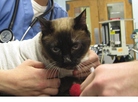
When Betty Gardner of Flowery Branch, GA, noticed that her 16-year- old Seal Point Siamese cat, Sissy, was confused and circling compulsively, her local veterinarian referred her to the UGA College of Veterinary Medicine teaching hospital.
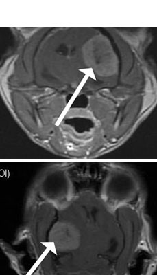
Highly detailed MR images taken at the BIRC revealed a large mass with characteristics common to a brain tumor called meningioma.
“We used the MRI to help guide our surgical approach,” said neurologist Dr. Simon Platt. “The cat is slowly recovering neurological function and doing extremely well. Without MRI, it would have been difficult to diagnose the lesion and impossible to operate safely.”
The new 3T MRI system may be able to pick up more subtle diseases and conditions, according to Platt. Its strength and functional capabilities give rise to new treatments for diseases common to animals and humans, such as strokes, brain tumors and even central nervous system inflammatory disease.
For comments or for information please e-mail: rcomm@uga.edu
To contact the webmaster please email: ovprweb@uga.edu
![]()