

How
a Slime Mold
Came to the Aid of
Alzheimer's Research
by Kathleen Cason
Intro/Proteins
run amuck
| The path to hirano bodies
Serendipity and beyond
|
What's ahead
Current students (bottom photo) are making new discoveries about what
Hirano bodies are made of and what they do in cells.
Front (l to r): Dong Huan Kim, Seonil Kim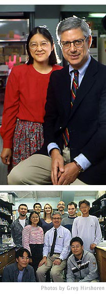 For UGA scientists Marcus Fechheimer (top right) and Ruth Furukawa (top
left), research is a team effort that includes graduate and undergraduate
students.
For UGA scientists Marcus Fechheimer (top right) and Ruth Furukawa (top
left), research is a team effort that includes graduate and undergraduate
students.
Middle (l to r): Ruth Furukawa, Marcus Fechheimer, Sang Deuk Ha
Back (l to r): Paul Griffin, Dan Del Portal, Heather Hensley, Rich
Davis, Patrick Stow (not pictured are Luke Hoagland and Caelin Ann Cubenas).
The path to hirano bodies
But how did a cell biologist studying cell movement and structure — in a lowly slime mold no less (see sidebar above) — end up investigating something that may have a role in degeneration of the human brain? It seems like a stretch at first.
Basic but complex questions in cell biology have piqued Fechheimer’s curiosity and driven his research program for more than two decades: How do cells move? What gives them shape? How do they eat?
The key to such riddles resides in a fibrous network inside cells, a kind of internal micro-scaffolding called the cytoskeleton. Besides acting like the “bones” of a cell, the cytoskeleton is an intracellular highway system, where specialized proteins scoot along the roadways toting organelles and other materials to the places they need to be.
“When we look at something that seems very obvious and beautiful like a crawling amoeba, it’s almost beguiling in its simplicity,” Fechheimer said. “But then you think about all the different molecular interactions and changes just in the actin when a cell is crawling forward. There are a lot of different things going on in different places. Figuring out all those dynamics is very complicated and very exciting. And we have great tools today to do it.”
To uncover the secrets of cell structure and movement Fechheimer’s team probed the inner workings of molecules that make up the cytoskeleton. The “roadways” consist of filaments made of three distinct proteins — one of which is actin — along with helper proteins that either anchor or bundle the filaments.
Actin doesn’t work alone. Among its helper molecules is a family of proteins that include one called the 34 kDa actin bundling protein. As its name implies, the 34 kDa protein packages actin filaments into bundles.
Actin bundling proteins play important roles in a variety of processes in the body.
“These actin bundles are ubiquitous,” Fechheimer said. For example, they have a role in hearing.
�The pressure wave that comes into your ear actually deflects actin bundles that are arranged like a staircase in the sensory hair cells and then an electrical signal gets sent to the brain,� he said.
During the past two decades, Fechheimer’s team has systematically answered many questions about the 34 kDa protein. They pinpointed where it is found in cells and what controls its ability to stick to actin. They’ve uncovered the protein’s structure. Along the way, they’ve looked at how it works by chopping the protein into pieces and testing how different snippets bind to actin.
To fine-tune their understanding of what the bundling protein does and how it works, they have altered the gene for the 34 kDa protein in small ways and inserted the altered version back into cells to see what happens.
That's what Andrew Maselli was doing when he discovered something else.
Intro/Proteins
run amuck
| The path to hirano bodies
Serendipity and beyond | What's
ahead
For comments or for information please e-mail the editor: jbp@ovpr.uga.edu
To contact the webmaster please email: ovprweb@uga.edu
![]()